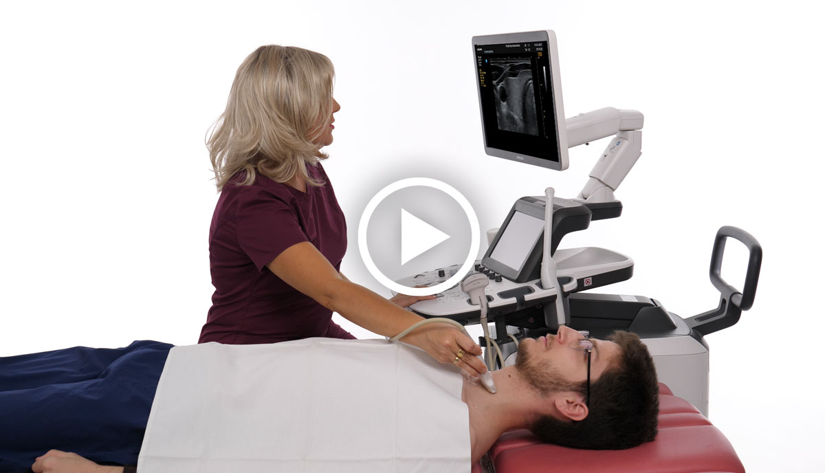
Ultrasound Diagnostics of Joints and Soft Tissues
Joint and soft tissue ultrasound is a non-invasive medical procedure that uses high-frequency ultrasound waves to create images of the inside of joints and soft tissues.
This technique is often used to detect inflammation or fluid in a joint, to detect early changes in joints, tendons, ligaments, muscles, and other soft tissues, or to detect various rheumatic diseases.
Ultrasound provides a clear visualization of the structure of joints, tendons, ligaments, muscles and other soft tissues.

This method uses high-frequency ultrasound waves to obtain an image of the organ and is often recommended as a preventive measure.
Ultrasound waves penetrate tissues and reflect off structures inside the body, creating an image on a screen. Based on these images, the doctor can examine joints, tendons, ligaments, muscles, and other structures to diagnose damage, cysts, or soft tissue tumors.
Who are the courses and trainings intended for?
Who are the courses and trainings intended for?
• General practitioners, residents, specialists, and subspecialists in various medical fields who want to gain basic and advanced knowledge and skills for independent performance of ultrasound and color Doppler diagnostics.
• Doctors trained to perform ultrasound and color Doppler examinations but unable to conduct a sufficient number of ultrasound examinations in daily practice to stay trained for routine ultrasound examinations.
• Experienced doctors who want to refresh their knowledge, receive a certificate, and earn points for license renewal.

